Anticuerpo IHC -- IgG4
Control positivo:
TonsilLocalización Celular:
CytoplasmSolicitud:
IHC-PAnticuerpo secundario:
iVision™Especificación/ml:
1、3、6、0.2(Concentrated)【Nombre del producto 】
Anticuerpo IHC -- IgG4
【 Especificación de embalaje 】
| Código | Clon | Especificaciones |
|
AR0518 |
SD413 |
0,1 ml , 0,2 ml , 1 ml, 1,5 ml , 3 ml , 6 ml , 11 ml , 30 ml |
|
AM0359 |
MRQ-44 |
0,1 ml , 0,2 ml , 1 ml, 1,5 ml , 3 ml , 6 ml , 11 ml , 30 ml |
【Uso previsto 】
Solo para uso de investigación. El reactivo de anticuerpo IgG4 está diseñado para identificar cualitativamente IgG4 mediante microscopía en secciones de tejido FFPE utilizando un sistema de detección inmunohistoquímico .
【P rincipio 】
La enfermedad esclerosante relacionada con IgG4 ha sido reconocida como una entidad de enfermedad sistémica caracterizada por niveles elevados de IgG4 e infiltración linfoplasmocitaria difusa con la presencia de muchas células plasmáticas positivas para IgG4. Las manifestaciones clínicas son evidentes en el páncreas, conducto biliar, vesícula biliar, glándula lagrimal, glándula salival, retroperitoneo, riñón, pulmón, mama, tiroides y próstata. Los análisis inmunohistoquímicos de la enfermedad esclerosante relacionada con IgG4 muestran significativamente más células plasmáticas positivas para IgG4 que las normales en los tejidos afectados. Agregue el anticuerpo primario para unir el antígeno en las secciones de tejido y luego use el anticuerpo primario de unión del anticuerpo secundario marcado con HRP para formar el complejo anticuerpo secundario - anticuerpo primario - antígeno . Cuando se agrega la solución cromogénica DAB, la HRP reacciona con el sustrato enzimático para producir un producto de reacción insoluble de color marrón, lo que indica indirectamente la existencia del antígeno.
【Componentes principales 】
Inmunoglobulina , diluyente de anticuerpos
【Almacenamiento 】
Conservar a 2~8℃ durante 18 meses 。
【Requisitos de la muestra 】
Los tejidos FFPE generalmente se cortan en secciones tan delgadas como 3 ~5 μ m con un micrótomo. Estas secciones luego se montan en portaobjetos de vidrio que se recubren con un adhesivo tisular.
【Protocol】
1. Sample preparation:Deparaffinize the slides in xylene Ⅰ, Ⅱ, Ⅲ for 5 minutes;Transfer the slides once through 100%, 100%, 95%, 75% alcohols for 2 minutes respectively. Rinse slides with deionized water for 30 seconds.
2. Blocking:Block endogenous peroxidase activity by incubating sections in 3% H2O2 solution at room temperature for 5 minutes to block endogenous peroxidase activity. Rinse the slides with deionized water for 30 seconds.
3. Antigen retrieval:Heat the EDTA Antigen retrieval buffer to 100℃. Then place the slides in the boiled buffer and continue to heat for 15~20 min. Naturally cool down for 30 minutes. Rinse the sample with wash buffer.
4. Primary antibody incubation: Drain the slides. Add primary antibody to tissue, incubate at room temperature for 30 minutes. (use antibody diluent or PBS as control). Wash the slides in PBST for 2 times, 5 minutes for each time. If the Primary antibody is concentrated, please dilute it to RTU(ready to use) according to the information on packing.
5. Secondary antibody: Drain the slides. Add secondary antibody to tissue and incubate at room temperature for 20 minutes. Wash the slides in PBST for 2 times, 5 minutes for each time.
6. DAB:Drain the slides. Add DAB to the tissue and incubate at room temperature for 5 min. Rinse slides with deionized water.
7. Hematoxylin staining:Drain the slides. Add Hematoxylin to the tissue and incubate at room temperature for 5 minutes. Rinse slides with water. Use the acid solution for differentiation. Rinse slides with water.
8. Dehydrate:Dehydrate the slides in 75%, 95%, 100% alcohols for 2 minutes. Dry the slides. Cover stained tissue with a coverslip using mounting medium.
【Positive localization】
1. Positive localization: cytoplasm.
2. Positive control: tonsil.
【Precautions】
1. Please read the instruction carefully and become familiar with all components of the kit prior to use, Strictly follow the instruction during operation.
2. DO NOT use the kit or any kit component after their.
3. Only trained professionals can use this kit. Please wear suitable lab coat and disposable gloves while handling the reagents.
4. Avoid contact of skin, eyes and mucous membranes with the chemicals.
5. DO NOT pipet by mouth.
6. Unused reagents, used kit and waste must be disposed according to local regulations.
【Manufacturer】
Company Name: Xiamen Talent Biomedical Technology Co.,Ltd
Address: No.3rd and 4th Floors , Building B10, No. 2068 Wengjiao Road West, BioMedical Park, Haicang District, Xiamen City, 36100 China
Tel: +86 592 6315755
E-mail: tlsw@talentbiomedical.com
Website: www.talentbiomedical.com
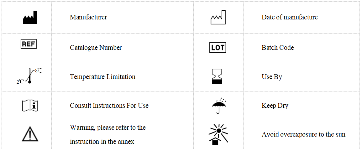
【Symbols】

Etiquetas relacionadas :