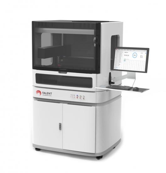
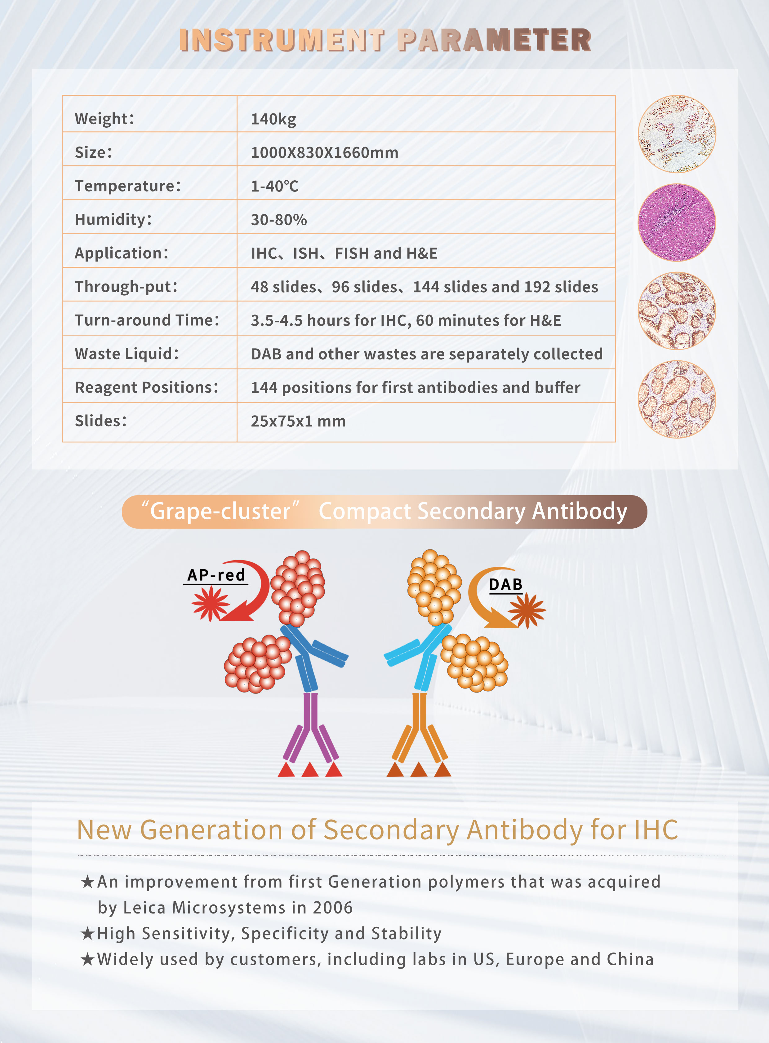
Peso:
170kgTamaño:
1000X830X1660mmTemperatura:
1-40℃Humedad:
30-80%Solicitud:
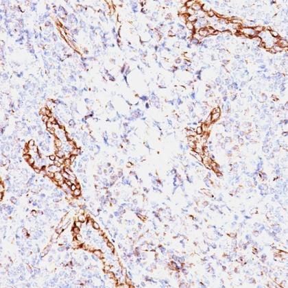
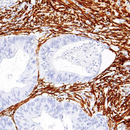
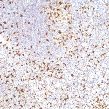
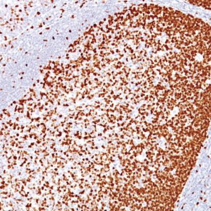
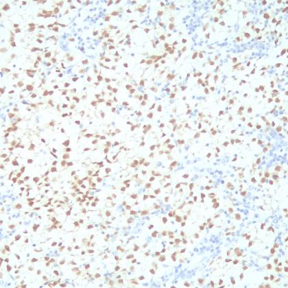
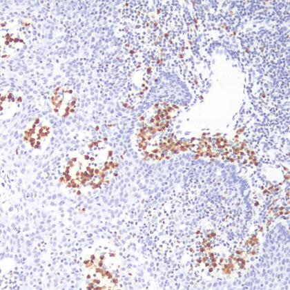
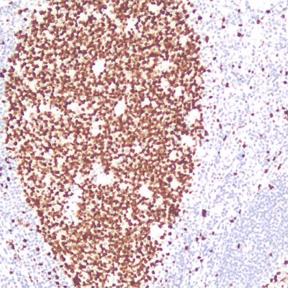
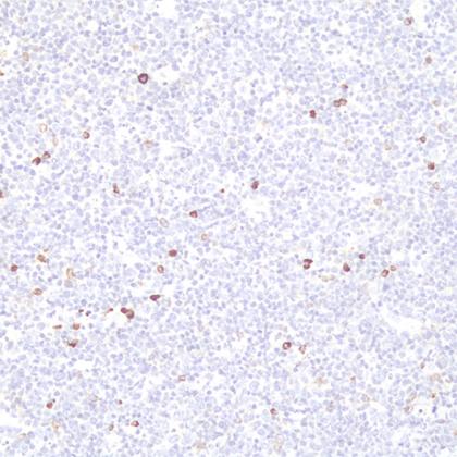
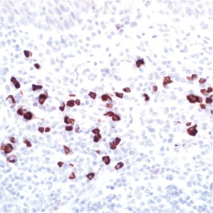
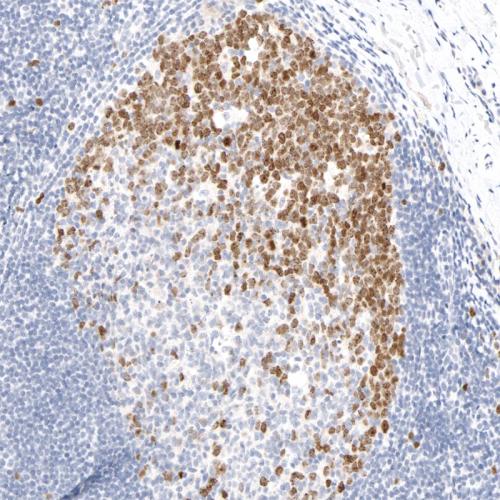
IHC, double-HC and ISH stainingRendimiento:
48 slides、96 slides、and 144 slidesTiempo de respuesta:
3.5 hours per roundLíquido residual:
DAB and other wastes are separately collectedPosiciones de reactivos:
144 positions for first antibodies and bufferDiapositivas:
25x75x1 mm

Etiquetas relacionadas :


Enlaces amistosos :
tianzuomedical© 2026 Xiamen Talent Biomedical Technology Co.,Ltd.Reservados todos los derechos.